Showing posts with label Embriology. Show all posts

Thursday, May 22, 2008

sheet 39 ::: the final one ::: edited :)

---The final anatomy sheet----
*In the name of Allah*

SPLEEN


What is the spleen ??
- its largest lymphoid gland in the body

Where is it?
It is located in the left hypochondriac region
Sheltered below left dum of diaphragm


It has three function:
1. storing your extra R.B.Cs
2.storing the old (worn-out) R.B.S
3. important for immunity.


Note: it is a completely intraperitoneal organ , except at its hilum.

Now let us have a glance on its parts :

It has the following parts :

2 surfaces : (1) convex diaphragmatic surface related to the left 9th , 10th , 11th Ribs
- with its axis parallel to left tenth rib -

) Concave visceral surface related to the stomach


2 Poles : (1) upper pole
(2) lower pole


please see page 2..

2 Borders : (1) superior border ( notched )
(2) inferior border ( sharp , smooth)


what about its arterial supply and venus drainage ???

Arterial supply ----> splenic artery (which is a branch from ciliac artery )

Venus drainage ----> Splenic vein ( which end at the portal vein )


Do know, my dear colleague ,that the Spleen follow the [ all or none ] rule !
That means that when its ruptured it should be removed , because if not it will cause a huge internal bleeding..


That’s enough about the spleen ….. lets talk briefly about pancreas


Pancreas


Little notes :
- acompound retroperitoneal endocrine and exocrine gland ( about 15 cm )
- comma-shaped extending from the 2nd of duodenum to the hilum of spleen
- located in the epigastric and left hypochondriac regions.


Has the following part :

1- HEAD : within concavity of the dudenum , received common and accessory pancreatic duct , bile duct course from behind !
2 -NECK : having the portal vein located behind it

please see page 3…..
3 - BODY : logest part , triangular in C.S splenic vein passing behind it
Splenic artery passing superior to it.

We said before that it is an endocrine and an exocrine gland at the same time.…..

Endocrine part : with in islets of langerhands : secreting 2 hormones :
1. Insulin : lowering the suger level in the blood
2. glucagon: elevating the suger level in the blood
Exocrine part :pancreatic duct : secrete to 2nd part of dudenum by :
- main pancreatic duct - accessory pancereatic duct

4 - TAIL : located at the hilum of spleen


good note : in future when you have a patient you should lay him/her on his/her back this is called “ supine ( recompetent) position “
**********************************


First you should all meet Nerdoosh ! He is an expert at embryology and he is helping us out with the last embryo sheet !!


Nerdoosh : before I start I feel I should straighten out a couple of things !

1-Blast : Original
2-Cyst: Sac
1+2 = BLASTOYCYST = ORIGINAL SAC

3-Mere : Subdivision
1+3= Blastomere = ORIGINAL SUBDIVISION


And with this my lecture shall begin !

This is a Continuation of the series of events of day 13 :

By the end of the second week the blastocyst is completely implanted and the endometrial implantation defect is healed .

Bleeding might occur occasionally !

Nerdoosh : it is around the 28th day of the lady's menstrual cycle !
14, ovulation day + ~ 13 days of pregnancy = 28!!!!!


Cellular columns from Cytotrophoblast invade the Syncytiotrophoblast forming the Primary Villi which is the beginning of placenta.

** I think you should start concentrating! New stuff starts here!


Primary villi will form secondary villi and then tertiary villi which is the final form of placenta.

At this time the intercommunication between the embryo – embryonic blood to be specific – and its mother's blood starts . However ,this communication is not complete ! Both are separated by a highly selectively permeable membrane.

*Nerdoosh : This permeable membrane is selective ! Not all that comes into mommy's blood will pass through… Nutrients are an exception and unfortunately so are the viral particles of measles and AIDS.

** :) If you guys just take a look at the hand out of this lecture you will notice something called "Connecting stalk" , this stalk forms the umbilical cord later on ! :) **


Once the embryo enters its third week , at day 15 , Gastrulation occurs .

Nerdoosh : Gastrulation is a process during which the morphology of the embryo is restructured by cell migration . It will change from a Bilaminar embryonic disk into a Trilaminar embryonic disk.

** You might want to take a look at your hand outs :) **

*Rapid development marks the beginning of the embryonic period : 3rd week – 8th week

*********************
Gastrulation explained!
1-At the beginning of the 3rd week a narrow line called the primitive streak is formed at the middle of the still Bilaminar embryonic disk.
2-Epiblast from the periphery will migrate towards the primitive streak, forming an elevation or a groove called "primitive groove".
3- Some of the migrating epiblast cells will migrate downward in the groove forming mesoderm ( it is between epiblast and hypoblast )
4-some will go down further to replace the hypoblast and give endoderm
5- Remaining cells will replace the epiblast and form the ectoderm. And with this the embryo is Trilaminar :)

Nardoosh: at this point I think we all arrived at the same conclusion! All body layers are derived from the epiblast!!

· Each layer is responsible for the formation of certain organs during the embryonic development.
1- Ectoderm: Nervous system + Epidermis of the skin.
2- Endoderm: Endothelial lining of digestive system, respiratory system etc.
3- Mesoderm: muscles, tendons, ligaments, bones, cartilage & blood vessels !


Nardoosh: Mesoderm is obviously the most important layer of all 3 layers ( AKA: germ layers ) .

With this I shall bid you farewell and leave room for Wa3d's and 3attili's longest dedication ever! Those 2 are breaking the record of the longest dedication written in the history of sheet writing if there is such a thing !


We, wa3d w 3abdelra7man , hope that we introduced a understandable and easy-to-study sheet :)


We feel obliged to thank Dr. Maher Hadidi for all the effort he exerted in teaching this course :)

We also wish you people the best grades in this course and future ones … Inshallah we will all make amazing doctors in the future ! Just remember: If you are going through hell , keep going on ! ** WE DIDN'T MAKE IT UP , SUM1 REALLY SAID THAT ! **


AND Finally we want to dedicate this sheet to our beloved friends and to all our colleagues in this faculty……

Shu r2yak 3attili nebda bel ladies be ma eno ladies first ?

Zein Najada ( eta oshkot ) , Haya Qudah ( finally a dedication … Yeppieeeee w 3attili b7gezlik el marah ele jay ! ) , Jamila 7iesat ( a7la ta7yeh la sokan jomhoriet al sal6 al sha8i8a ) , Suzan Mbydeen ( sum people r still waiting 4 that mansaf gurl ) , As7ar Tarawna , ala2 7jazi , Shatha 3attili ( 3ala rasi che guevara ) Dania Dahmash , Bayan 5door,saja 3r3r , Suzan Momani , Ruba al mu7taseb , Lamoosh jamal , Sawsan tabaza , Sondos al 5ateeb , Elham Qudah (dedication ^2 :P) , Batoul 3toom , Nadine 3adayleh , , Dana mel7em Tamara Darweesh , raya 7alawani , magd 5adr, amal 7osban ,Manar Jwainat , Fara7 mashagbeh , 3abeer 5watra, samar 68a68a , muna talal ( 3al 3afyeh:P) , ala2 jabre ( el m63m mo elhm :) ) , dana 6awalbieh ,Samar al ra7meh , nisreen abu 3osbeh , rawan 3'azal , rasha jabra , ala2 3abid, jeelan , suzan al musa , Isra2 zawawi , dina 3ammari , aya bani kinana , rozana ziadat , asma , lana 7adadin , ruba halaseh, dina ramini , razan abu dieh , rawan ( lovek 4 taking psycho :D ) ,ala2 7laisi, aseel abu shanab , du3a2 jara7 , du3a2 abu jame3, aseel al sayed ,dima , nadia ,rawan sale7,nuha, ,yasmeen el so5on , anne nimri !!

And with no further delay, the guys :

Ahmed el isa ,(abuel3ss) , ,fr7an el kooz ( jovial ) ,) wa2el 6o8an (alf mabrook:P) , ayyoub (yellow smile ), , 5alid 7gair (4-2-4) , m7md abu hanieh ( abu gor3a ;) , w aseem samara ( kasha6a),shaker borhm , hammam realat , m7md shawaf ( 3a rasi kol el i6allieh )fadi halaseh, yezan 3tom( ml7 3l sala6a) , martin qaqesh(kef el shbab) , bashar el rama7i , 3omar el lozzi ( neshfat el mae) , nidal matani (no visa,no league, no thing :D), 3’aleb 5rfan , 7amzeh 9araiera , m2moon sha3ban (ma bnnsak) , yazeed neef , abu 7alaweh , mo2ayad kettane ,sami 3abdeen , a7mad 3abed, 3amer 7ajaj,m7md saree3 , m7md maslamani , Bandar maslamani, 7amzeh jassar ( elsalam la allah ;p) , m7md el sboo3 , 7amzeh 9bai7 , , rami yagi, hashim abu m7fooz, zain rostom ( 3ala rasi walla) , nemer sh5shier , 3mr el 5alili , 3esa 38el , zaid 8ndeel, 3omar dodeen , bashar sharma ,7anna barghoth , tamimi ,8osai abu 3azzam , zaidon , 3bdelr7man el 56eeb , m7md 3’naem (pre-sheet), mostafa aburahmeh , a7mad abu 5adegieh, , m7md 8awasme , abu qamar , abu d3aig ( allah be3en 3l basa6 ), a7mad shalabe , el shareef , 3omar radaiedeh , yazan radaideh ( m5drat ) , , laith 7addad , osama , yazeeed 8aesieh , waleed 8adre , osama farrogeh ( farrogte) , mohanned ( ya 7abibi la t3eed wrae) ,A7mad 7orani ( hay dedication 3ashan ma t7ke 3an el sheet tb3tna!)


Ma azon nsena 7ada… :P

Wa 5etamoha mesk m3 wa3d Sweilmyeen w 3bdelra7man 3attili :D ….
Best wishes..

---WE LOVE YOU ALL, THANK YOU ALL---


Note:- organic final exam will be from (3:00-5:00) PM
-islamic culture final exam will be on 4/6/2008 at 11:30 in IT


** Corrections are in purple bold...becareful !!


Tuesday, May 20, 2008

sheet no.37

Notes for the last lecture:

-Outer cell (trophoblast) mass is nutritious.

-blastocyst cavity is the pool that allows the embryo to grow the way he wants freely.


Implantation


-Pregnancy starts when the Blastocyst loosely attached to the endometrium at Day 6(from fertilization)---->anchored implantation.

-Trophoblastic cells above inner cell mass attached to uterine wall by invading its epithelium and underlying stroma.


-At Day 7 implanted Blastocyst interrupted the regular sexual cycle affected by hCG hormone production from trophoblastic cells of the implanted Blastocyst.

*hCG---> (human Chorionic Gonadotropin)

-Chorion: Fetal portion of placenta.

-Gonad : sex organ (ovary here).

This hormone prevents degeneration of corpus luteum (C.L.)--->in the first 3 months, and maintains it as C.L. of pregnancy.

*Remember that C.L. will degenerate at Day 28 (from ovulation) and become corpus Albicans if no fertilization occurs.

-So we can detect pregnancy at Day 7.


At Day 8:

The trophoblastic cells differentiate into 2 layers:

Inner layer: Cytotrophoblast (mononucleated distinct cells).
Outer layer: Syncytiotrophoblast (multinucleated protoplasmic mass lacking cells) which is highly energetic.
Both 2 layers become part of the chorion.


The inner cell mass (Embryoblast) differentiates into 2 layers:

1- Hypoblast (with cuboidal cells) àendoderm to be.

2-Epiblast (with columnar cells) àectoderm to be.

Both layers form the Bilaminar embryonic disc.

A small cavity appears within epiblast then enlarges to be the Amniotic cavity.

Notes:

-The embryo has 3 layers:

Ectoderm (outermost).
Mesoderm (middle).
Endoderm (innermost).


-In Day 8 it will forms 2 layers & 2 cavities (Weak 2 loves no. 2).

-Until now there is no complete implantation.


Day 9:


Notice that:

-Syncytiotrophoblast will spread around(beldozer).

-Blastocyt embedded deeply in endometrium.

-Sycytium show small fluid-filled cavities (vacuoles)àfuseàto form trophoblastic lacunae.

* Lacunae mean---->cavities / small lakes.


Days 11 & 12:---->complete implantation


Trophoblastic lacunae fused to form lacunar network which communicate with maternal sinusoids to establish the Uteroplacental circulation which is the first communication between the mother & the embryo.


Day 13:


It has 3 main events:

Endometrial implantation defect healed.
Bleeding occasionally occurs at implantation site at Day 28 (from ovulation) of normal cycle?
*The bleeding may fool the lady and make her think that she is having her period.


Cellular columns from Cytotrophoblast invading Syncytiotrophoblast forming Primary Villi which is the beginning of placenta.
Cytoplast will form finger like pillar & penetrate inside forming Primary Villi.

*Now we have completely implantation (implantation ends at day 13).


Note: Fertilization occurs at Day 13 of ovulation so when we want to do the math we add 13.

e.g.: Day 8 from fertilization = (8+13=21) Day 21 from ovulationàsecretory phase of uterine cycle.


-so by day28(13 from fertilization) the embryo differentiates into 2 layers(endoderm-ectoderm) and placenta will have started then.


"Two little mice fell in a bucket of cream. The first mouse quickly gave up and drowned. The second mouse, wouldn't quit. He struggled so hard that eventually he churned that cream into butter and crawled out. As of this moment, I am that second mouse."


I want to dedicate this sheet to:

Children of Palestine, Iraq & Lebanon (You are the real men).


And to all my friends who:

-Study on medicine faculty:

5aled 7ajeer(Mr.”bla 10 3la al shagarah”),Ebraheem Abo Dya, M2moon sha3ban, Ahmad Abo Hallawah, Osamah 5aleel, Ahmad Abo Khadeegah ,5aled Daye3, Yazeed Al-naif , Yazeed Qaisyah , M2yad Ketaneh,7amzeh jassar , Qosay 3zzam ,Moh. Tamimy, Moh. Momany , Yazan 3toom(y36eek el 3afyeh) ,3dnan Al-3aref (ya 7ram Chelsea) , mo2men K7aleh , Abd Allah Tayyem ,Ebraheem Al-Sa3eedy , Moh. Al-quasmeh ,Moh. Abo 3rjah, Ahmad 7ra7shah , Ahmad 7na6y , 9ohayb Al-3bady , samy 3bdeen , Ahmad Al-shalabi, Moh sbo3, Zaid mal7ees .

-Study on other faculties:

Sa2ed kharoob, Moh. Bdar, J3fary (The tall) ,Ahmad (Sokkar) , Mahmood & ahmad Abo Znai6, Monther, Ahmad hamshary , 6ohayb, shukri, 30mar semreen,Wael 3lawneh,Muhannad,Nusaiba .

-Study on other Universities:

3la2 mara3’y, Wael Abo Hassan, Fat7y, Moh. Faroq, 7osam.

-Study on other countries:

Abo hmade (King kong) , Marwan 5amees.((In Egypt)).

J3fary”the short” (sht2nalak wala) ((In U.S.A.)).

-And to Dr. Maher Al-Hadidi.

-And to my little brother Hashem Al-Faqih.


*And to people who are special to my heart*

Mohammad Al-Faqih

Wednesday, May 7, 2008

sheet no.34

At ovulation (day 14) the secondary oocyte usually come from the ovary due to the effect of surge (large amount) of luteinizing hormone, at that time it is ready for fertilization.
Each ovulated secondary oocyte at that time is covered by 2 layers :-
-outer layer (corona radiata) which is part of the follicular cells.
-inner layer (zona pellucida) a transparent layer which is produced by the outer follicular cells and composed of glycoprotein.
(the secondary oocyte is in 2nd meiotic stage)


Fertilization:


A process by which haploid male gamete fuse with female gamete to give single diploid nucleus.
Normally occurs 12-24 hours after ovulation while the ovum in the upper part of uterine tube.
N.B
(-only 300-500 sperms out of 300 million can reach ampulla where only one of these fertilize the oocyte
-the trip from cervix to oviduct needs 2-7 hours)
Arrived sperms are unable to fertilize oocyte except after 7 hours of conditioning in female genital tract for
1. capacitation: epithelial interaction between sperms and mucosa.
Only capacitated sperms can enter corona radiata and undergo acrosomal reaction
2. acrosome reaction:
Acrosome which is located at the head of sperm enter the ovum in 3 steps:
*penetration of corona radiata :( not all of sperms enter the corona radiata but only many of them enter) by entering the corona radiata, corona radiata will change interaction with other sperm
*penetration of the zona pellucida: the acrosome will secrete digestive enzyme locate in the acrosome which called acrosomal enzyme specially acrosin which is received by receptors on zona pellucida called ZP3 glycoprotein which act as sperm receptors that trigger acrosin to digest a path through zona pellucida.
*fusion of membranes: after gametes are contacted membranes of gametes fused, head and tail of the sperm penetrated into the cytoplasm
Oocyte response
1- cortical and zona reaction:-depolarization of oocyte membrane prevent poly spermy(entering of more than one sperm) -inactive ZP3
2-resumption of 2nd meiotic division: female pronucleus + 2nd polar body
3-meta bolic activation of oocyte
Result of fertilization:
1-restore diploid number of chromosomes
2-stimulate resuming 2nd meiotic division
3- determines the genetic sex of the new individual
4-initiates cleavage about 24 hours post-fertilization


After fertilization of secondary oocyte it will form female pronucleus and there will be a male pronucleus

Male pronucleus will approximate female and sharing same membrane and chromosome will intermediate (the DNA will duplicate) , then the 2 pronucleii fused forming onr membrane (one nucleus) with male and female chromosome which will aline at equator , then division will occur to give two-cell stage.

Morula: a ball of cells appearing from outside and (the number of cells is 16 at the early morula but it will become 32 at late marula) it will start entering the uterine tube.
At day 6 it will wander inside uterus
(N.B the uterus at that time is in secretary stage under control of progesterone)
When arrive this area the secretion will enter inside the marula
The fluid of uterine will enter between the marular cells separating it into:
-inner cell mass (Embryoblast) which give the embryo
-outer cell mass (trophoblast)
-cavity

At this stage the morula is ready for implantation.

To all my friends specially:

A7md 2bo-7lawe, yazeed al-naif, 5aled 7ajjir, mo2ayyad kettane ,yazeed qayseye, osama 5aleel, qusai al 3azzam, a7md 2bo 5deje, a7md al-shalabi,
Amjd 3yyash (el 8a2ed el ramz), 7amze jassar,m7md al faqeeh, 3omar el 7aj,
Yazan 3toom, nidal matani, m7md qteshat(mesh naseek ylle nasene), m7md sboo3, a7md 7ra7she, 9ohaib el 3badi, ibrahim 2bo deyye ,ibrahim el 93idi, hmmam ryalat, 7azem, m7md tyseer, sami 3abdeen, a7md el 3esa, 3bd el ra7man 3attili, mo3ath el zo3bi, martin qaqeesh, 3'aleb 5erfan, m7md bdr,
m7md 2bo haneye, zaid qandeel, 3amer 7jaj, 3omar el-lozi, mo2men k7aleh, m7md qawasmeh
w slam 5a9 la el shbab msh el 6eb:
Muhannad bdair(Hannood)
Wa2el 3lawne
Nusaiba el jalodi
3omar smrain
Special thanks to yazan 3toom

****With best wishes from
Ma2moun sha3ban

"don't limit your challenges ,but challenge your limits"

Wednesday, April 30, 2008

sheet no. 31

Notes about oogenesis


* Follicular cells: for maturation (maintenance, protection) of oocyte.


* Corpus Luteum (the yellow body): -It collects fat that’s why it's yellow in color

- Its center has blood


*Corpus albicans (the white body): It's fibrous tissue (useless)


**The last line in oogenesis's handout was missed, the whole sentence is:

(Ovulatory Stage: At ovulation 2ry oocyte enter Meiosis II but arrested in Metaphase, only completed at fertilization to five mature ovam otherwise, it degenerate after 24 hours)


Female Reproductive Cycle


*It's a continuous monthly cycle from day 1 of Puberty till day 1 of menopause.


*The ovary is not controlled by it self it's controlled by Hypothalamus, pituitary gland (they are controlling the ovary and the ovary will control the uterus).


*Female Reproductive Cycle involves Hypothalamus, pituitary gland, ovaries and uterus. It includes monthly cyclic changes in structure and function of ovaries and uterus in preparation for pregnancy.


*At puberty hypothalamus secretes Gonadotropin Releasing hormone (= GnRH) that give the command to Anterior Pituitary gland to secrete 2 hormones:

1- Follicle stimulating hormone (FSH)

2- Luteinizing hormone (LH)


Note that (Gonadotropin) =

(Gonado –) it means: Main sexual organs "In male they are testis and in female they are the ovaries)

(- tropin) it means stimulating


*Female Reproductive Cycle is divided into 2 cycles:

1- Ovarian cycle

2- Uterine cycle


Ovarian cycle

FSH & LH stimulate and control cyclic changes in ovaries (it affect the ovary by)

Growth and maturation of follicles (Proliferation of follicular cells)
Ovulation
Corpus Luteum


Uterine cycle

Ovarian hormones control monthly cyclic changes in the endometrium which involve 3 phases:

Menstrual phase ( Bleeding phase )
Proliferative phase
Secretory phase


*The typical cycle in female is 28 days starting by day 1 of bleeding (menstruation) till day 1 of next bleeding.


* The typical mid time of it is (day 14) which is ovulation time


* We can divide uterine cycle into 2 half's:

1- The first half is before day 14 (from day 1 to day 13)

2- The second half is after day 14 (from day 14 to day 28)


Uterus

The endometrium of uterus consist of 2 layers (from in to out ) :

Functional layer : it's formed of 2 parts ( compact layer , spongy layer ) because it's the layer that discarded ( removed ) every month .
Basal layer : Regenerative layer which regenerate formation of discarded functional layer .


* Uterine cycle controlling the structure and function of uterus specially endometrium by hormones released from the follicles or corpus Lutem.


Proliferative phase of uterine cycle


i.e.: regenerating (replacing) the discarded functional layer by basal layer which is controlled by ( Estrogen hormone ) secreted by follicle cells during the first 13 days .

That's why the first half of uterine cycle is controlled by Estrogen


* In Proliferative phase :


1- The epithelium is thicken 3 times

2- Glands will form

3- Arteries will reform ( repair )


About day 13 or 14 Luteinizing hormones will be secreted in large amount ( a surge of LH ) affecting the follicle which will rupture and burst to expel ( let go of ) 2ry oocyte at day 14 .


**at this time the epithelium is replaced to become 3 times in thickness, blood vessels will be repaired, glands enlarge, but uterus is still not 100% ready for implantation.


Secretory phase of uterine cycle


* After day 14 Corpus Luteum will take over of 2nd half of uterine cycle ( from day 14 to 28 ) by secretion of Progesterone .


Note that ( Progesterone ) =

(pro-) it means (with)

(gest-) it means (pregnancy)

(-on) it means (Formation)


In Secretory phase :


Progesterone will structure gland to swell and secrete more fluids
Blood vessels enlarge and congested with blood and will spiral
Secretion will be increased
The endometrium is ready for implantation ( healthy media )


* At day 28 if fertilization doesn't occur Corpus Luteum will degenerate and its cell will not secrete Progesterone è lower level of this hormone in blood will cause the blood vessels to leak blood from their wall or arteries will be constructed causing Ischemia ( Death of epithelium ) è there after blood will be collecting below epithelium and will be (sluft = يقشر) out within uterine cavity and will go out .

* By finish of day 28 the bleeding starts .

*blood amount in normal is80ml


Contraceptive Pills


* They mainly depend either on Progesterone or Estrogen by inhibiting or play games around ovulation


* If we don't want ovulation to occur we give Estrogen in the first half of the period è the level of Estrogen will raise and so it gives feed back to FSH and they will not secrete it ( False message )


* If we want pregnancy ( A lady with a problem ) è we give her FSH


I want to dedicate this sheet to all my colleges in our faculty specially to those who left their country and came to Jordan to study medicine, by the name of all Jordanians we say " Welcome to Jordan , it's your country so as it's our country, we all are one nation " , and they are :


From Syria : Khaled Daye3 , Moh'd Fadel


From Palestine : Moh'd Abo Haneyeh , Bolos Mansor , Waseem samara , Moh'd 3awwad , Rand Abo Lebda , Aseel Hoso , Mahmod Shalabi , Ayham Saleh


From Yemen : Mokhtar Al Warafe , Moh'd Shaye3


From Iraq : Fahed Adel , Moh'd Wazeer , Shahed Thaer , Sa7ar Basel , Senan Adnan , Bashar Yousef , Bashar Saba7 , Maryem .

And To whom I forgot …..


Special thanks to Our colleges who built the website for the sheets

( Wa3ed Swelmeen , Wael Tokan, hamza jassar)


Finally , A7la ta7eyeh men shabab el 6eb la morash7na el saneh el jay …. Arw3 shabab el dof3a ****( Ghaleb Kherfan )****

Tuesday, April 15, 2008

sheet no. 23

By the Name of ALLAH


Thorax

Thorax is the part of the body that extends from the root of the neck to the Diaphragm.
It has the following parts :
1-Skeleton which called (Thoracic cage)
2-Coverings which called (Thoracic wall) .
3-Toracic cavity And contents.
Thorax has inlet (Thoracic inlet) which is narrow and outlet(Thoracic outlet) which is wide covered by the Diaphragm that separates thoracic cavity from the abdominal cavity.

*Thorax is convex at all sides.


Coverings
1-Skin
2-Superficial fascia
3-Deep fascia
4-Three layers of muscles (Three intercostal muscles)---->soft structure
5-Ribs--->hard structure
6-Pleura( الغشاء البلوري للرئة ) which is a serous membrane containing fibrous tissue and proteins , this membrane in cold conditions become loose so (you can not breath)Because this pleura is highly elastic but in cold conditions it descended so the nerves would be tight causing a severe pain and can not complete the respiration this called (pleurisy).


Thoracic cage
Consist of:

1-Sternum (anterior).
2- Twelve thoracic vertebrae(T1-T12) (posterior)
3-Twelve pairs of ribs on both sides.

**Sternum (breast bone) it is like a knife or more specifically like a dagger So it has three parts:
1-Handle (Manubrium)
2-Body.
3-Tip which called(Xiphoid process)
**The Sternum is a flat bone with spongy tissue .

Manubrium Articulates with the first Rib and a half of the second rib.

Body Articulates with half of the second rib to the seventh rib.

Xiphoid process It is a hyaline cartilage And ossified after the age of 40
And before this age it is cartilaginous

Sternal angle(angle of Louis)

-It is located between The manubrium and body, it is at rib two level
- It is a good clinical land mark That marks the followings:
1-Rib number two to count ribs .
2-Biforkation site ( منطقة انقسام) of trachea .
3-Aoartic arch.
4-Inter vertebral disk between T4 and T5.
*Body of the sternum located anterior to the heart so in C.P.R(Cardio pulmonary resuscitation) we compress the heart between sternum and vertebral column.


[Embryo notes for the last lecture]


*Gonia = off spring (ذريِِِة) .
*Zoon = life.
*testis are outside but they form in the posterior abdominal wall then migrated out, why? Because they will not do its function on the body temperature
* Immigrated testis called undescended testical
* primordial germ cells arrive testis in the fifth week and start to enlarge(multiply) themselves by mitosis.
*Spermatogonia stay in rest until puberty (in males) but oogonia in females stay in rest until (birth) .
*Type B cells are committed to continue the course (fertilization).
*Multiplication started before birth
*spermatogonia and spermatocytes(1ry-2ry) are 2n the continue until they give 1n which is the sperm.
*Maturation begins at puberty.
*Spermatocyte cells are commited for formation of sperms
*Early spermatids are immature sperms.
*inactive=resting=dormantàlike dipluteen stage in females.
*spermatogonia type B is the which will continue the course.
*sex cords:àprimitive(without lumen)(before puberty)
àseminiferous(with lumen)(at puberty)


*some correction for sheet 22

1-Supplied by peronial artery , branch of posterior tibial artery
2-propulsive force by all muscles of the posterior comp. of the leg but the most powerful two are gastocnemius and musclessoleus
3- Tom has very nice doga and pigs >>>the muscles named from medially to laterally.


I want to dedicate this sheet to all my colleges in our faculty specially to those who are from al-ittihad school and they are:

Ahmad al-harahsha(captain), qusai al-azam(az az ya toto), yezan 3toom(wa7sh el-anatomy ), Ibrahim al-saeede(safe area!!) . mohammed al faqeeh , mohammed al tameme, abdallah tayem.
Ruba al-mohtaseb ,Do3a Al-jarah.Bayan al-khdoor ,Shatha al-attili ,Sondos al-khateeb , lama jamal , Rawan ghazal, Suzan mbaydeen , Rawan saleh,ro2a sameeh , 2la2 hijazee , Do3a abu jam3.

And to those who are special to me :

Adnan el-3aref(cute skalamboooo) , mo2men kahaleh , zaid malhees (el za3eem) , hazem abu 3sabeh , Omar el-haj omar, Yazeed al-neef(my heart),***Mo2ayad kettaneh***
Ma2moon shaban (snow white) , ahmad abu-halaweh.

And to those whom I Knew them in the university:

Sawsan tabaza , jeelan ,Dana mel7em, wa3d swaylmeen ,Fadi halsa ,Sali salman, mohammed ryalat ,mohammed qtayshat , mokhtaar,mo0hammed bader . mohammed abu haneh , ayham. ,waseem , shaker ,.

And please forgive me those whom I forgot .

Your college Ahmed abu-khadigh

Special thanks to my brother yezan 3toom

A lot thanks to our brilliant doctor :Dr. maher al-hadidi

Saturday, April 5, 2008

Some notes on the last Embrio lecture ....


1-no pregnancy occurs--->carpus luteum will shrink until degenerateàprogesterone levels will go down--->C.luteum will be converted into C.albicans(nearly at day 27)àendometrium replaced by myometrium---> second period will start.

2-follicle:ovum surrounded by nourishing cells

3-estrogen--->first 14 days of period-->before ovulation
Progesterone--->second 14 days of period--->after ovulation

4-carpus luteum is formed by remains of 2ry oocyte after ovulation

5-carpus luteum is responsible for:
-second phase of female period
-pregnancy
-progesterone secretions

6-ovary forms before 5th week

7-ovulation(of 2ry oocyte) occurs at day 14

8-ovarian layers after modified peritoneum are:
-Germinal epithelium
-Tunica albuginea
-ovarian cortical region(cortex of ovary)
-medulla

9-hilum:receives arterial blood supply and sends Venus drainage to and from ovary--->by ovarian blood vessels

10-follicles--->primitive(primordial)
---->growing (secondary)

Friday, March 21, 2008

sheet no.16

Cell Cycle


Consists of 2 major events:


a) Interphase.
b) Mitosis.


- Interphase is longer than Mitosis.


- In the Interphase the cell grows and duplicates its organelles and genetic material.
The Interphase stage consists of:
1) G1 phase: this phase is the major period of cell growth during its lifespan.
2) S phase: (synthesis) is the phase in which the cell duplicates its DNA.
3) G2 phase: is the phase in which the cell undergoes a period of rapid growth to prepare for mitosis.
M = Mitosis Stage
I = Interphase stage
- G0 phase: Some cells (like muscle cells) don’t divide more after mitosis and they become in a resting stage. Peripheral lymphocytes are stimulated to re-enter the cell cycle and divide after exposure to radiation, wounds, or diseases.
- Some cells leave the cell cycle(rest) permanently (like muscle cells(myocytes) and nerve cells(neurons)) while others leave it(rest) temporarily (like peripheral lymphocytes).

-In case of bone fracture:
a) Periosteum: is a membrane that lines the outer surface of all bones and it contains osteoprogenitor cells which produce osteoblasts. Periosteum is stimulated to produce osteoblasts when the bone is fractured.
b) Osteoblast: is the matrix formation of cell. It produces osteocytes.
c) Osteocyte: are maintenance cells.
d) Osteoclast: remodeling cells to reshape the fractured bones.

- How Cells Divide:


1) Somatic Cells – by Mitosisàgive 2 identical daughter cells
2) Reproductive Cells – by Meiosisàgive 2 in identical cells


Tuesday, March 11, 2008

Sheet no. 13

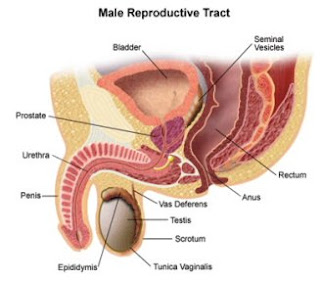
Male Genital system :


Parts :


1)Two testis
2)Two vas deferens(ductus deferens)
3)Two epididymis
4)Two seminal vesicles
5)One prostate
6)One urethra


Testis: [1]
-Paired organs dedicated to produce the male hormone called Testosterone and spermatozoa(sperms).


-2cm wide/3cm thick/4cm long.


-Ovoid organs located within pouch of skin called Scrotum outside the body.


-To functionàthe testis does not function on body temperature(37 degree) but on 3 degrees below it(34 degree) for production of sperms to happen otherwise they will not function at all.


-That is why if testis does not migrate from it's original position (normally)which is located at posterior abdominal wall(the same as ovary)àwe have to descend it down to it's functional position(outside body) by surgical operation.


N.B: the ovary function inside the body at it's temperature(37 degree) unlikely to testis.


-Clinical Note: if the fetus has completed the full term(9 months) and testis or ovary remains in post.abd.wall then it must be descended down by surgical operation.
-testisàinside bodyà(zero-shaped) 0
àoutside bodyà(rounded)


***Testis is divided into parts called:
1-Testicular lobules:


>each is pyramidal in shape
>each testis contain 250 of them(for both 500)
>1-4 tubule in each
***each testicular lobule contain
2-Seminiferous tubules:
>highly coiled blind tube(blind tube: one way or direction tube)
>(30-70)cm long
>produce spermatozoa(sperms)
{spermatogenesis}
N.B: sperms move only in one direction inside S.t
***
Spermatogenesis (sperm production)start in male at puberty it ends nearly on age 60
Oomatogenisis (ovum production)start in female at birth
So it ends nearly on age 45.


[2]Epididyms


-Highly convoluted tubule
-6M in length(sperm will grow until maturation during passage through it)
-divided into head, body and tail
-it stores and matures sperms after being produced by testis


[3]Vas deferens


-long muscular tube
-45cm long
-It extends from tail of epididymis to ejaculatory duct close to seminal vesicle.
-It conveys mature sperms from…to…(mentioned in point 3).


[4]Seminal vesicle


-two horn-shaped organs
-on posterior surface of urinary bladder
-each form of 15cm coiled tube(not a sac)
-secrete Fructose important for sperm motility
-rich seminal fluid that constitute by 70% to ejaculate
-resemble testis where both contain tubes
-it is the only organ in body that secretes fructose so women doesn't secrete fructose at all.


N.B: Fructose is used in forensic medicine as evidence for sexual assault(rape).


N.B: our source of fructose is from plants.


[5]Prostate


-base above/apex below
-below urinary bladder
-pyramidal-shaped fibro muscular glandular organ(chestnut shaped)
-surrounds prostatic urethra that passes through it
-form of 5 lobes
-receive ejaculatory duct in urethra coming from vas deferens and seminal vesicle
-secrete alkaline fluid(alkaline phosphatase)to neutralize the vaginal acidic secretions
-secrete product that prevents infection


N.B: there must be two conditions for sperm motility
1)Fructose
2)Alkaline phosphatase
N.B: the contracepted pills prevent pregnancy either by
1)increase acidity of vagina(vagina dosh)
Or
2)decrease amount of progesterone/estrogen.


N.B: in both male and female the genital organs are posterior to urinary bladder and anterior to rectum with difference that female G.organs are larger because of uterus.


N.B: Aorta artery divides into right and left common iliac arteries and each of them gives also external and internal branches.
-internal: for pelvic organs and internal structures(U.B/Rectum/Vagina/prostate…etc).
-external: for lower limbs.

*************************************************
I'd like to dedicate my work to all my colleagues in faculty

16 days remaining for exam


God help us


With my best wishes


Yezan Atoom

Wednesday, March 5, 2008

sheet no. 10


Uterus:


Thicked wall pear-shaped muscular organ
Located between urinary bladder anterior and rectum posterior
Found in: -pelvis (non pregnant ladies)
-abdominal pelvic cavity (pregnant ladies)


Has the following parts :


Fundus:


Convex
Located above the entrance of fallopian tubes
It is tilting(bending) forward
Press urinary bladder<>


Body :


It is about 2 inch in length
Has a triangular cavity called uterine cavity with three openings
tow uterine tubes above
internal os of cervix below
has anterior wall and posterior wall
posterior wall is the common site for
implantation of fertilized ovum(zygote). That’s why a pregnant lady is advised to sleep on her back to avoid abortion in the first 3 months of pregnancy and to assure good implantation.
Anterior wall is not good site for implantation and in case of that pregnant lady is advised to sleep upside down.


Cervix:
The most sensitive part in the female genital system.
Cylindrical about one inch in length
Has the following parts (from out to in):


external opining(external os)
it is circular in a lady never delivered a baby (never pregnant) (nullipara)
it is horizontal in lady with multy pregnancy (multipara)
doctors measure the width of the external os by inserting their fingers
external os is important to know if the lady has been delivered a baby before or not


internal opening (internal os)
communicate the cervix with uterine body


cervical canal:
a spindle shaped canal
communicating vagina with uterine cavity between internal and external os
it is the part that dilate before birth
layers forming the walls of uterus :


1-inner layer (endometrium)
transitional epithelium
regenerated every month due to the period
all should removed for good implantation but sometimes part of this layer still exist causing bleeding . that’s why lady with this case her’s endometrium should be removed to build anew layer
the endometrium builds a lining periodically which, if no pregnancy occurs removed by lady’s body

2. myometrium: thickest layer

3.perimetrium : mostly is peritoneum (kind ofmembranes)


*********


Position of uterus:


Normally:
The uterus is a pelvic organ before pregnancy and abdominopelvic organ in pregnancy (that’s why the pregnant lady can easily be tired and cant lay in her back because baby press on anterior organs especially diaphragm)
The uterus is antiverted and antiflexed (bending forward on the cervix) touching the urinary bladder


Abnormally:
Uterus may be bending backward(retroflexed) and retroverted on itself compressing on rectum and that leads to severe low back pain or diarrhorea
Usually pregnancy will not occur leading to abortion
Ladies with this situation is advised to lay on her abdomen
Doctors can fix this situation by pulling uterus from two ligaments


The end


I want to dedicate this sheet to all my friends whom I love especially
Zaid el 7loo, 7anna bar3’ooth, zaid shmessanee, tamara darweesh, 7amzeh jssar,marten 8a8eesh,nidal matani(a7la arsenal….. ma3lesh 7anna mogamaleh),samee7 matani ,abo 86eesh(mohd shwaf)
, el shareef yoossf kettaneh ,3meer el 5aleele,wa2el 6oo8an,feraas nemer,mo2ayad keetaneh,basher el rama7i ,mohd 3abood, a7mad 3ababneh ,mo3ath el ze3be, mohd 3assaf,leena el gaberre-7aneen 5reees(mis 3la raseee wallah), ,hammam ryalat

By: hisham hirzallah


Picture by : Yezan 3toom

Wednesday, February 27, 2008

SHEET NO. 7

- We have 2 types of cells :
1. Somatic cells : myocytes / osteocytes / hepatocytes which grow in number by a division called by mitosis producing 2 equal daughter cells
2. Sex cells : in the ovary or testis divide using a process called meiosis in which a mother cell gives 2 none equal daughter cells

- In human cells we have 46 chromosomes each chromosome has a maternal part and maternal part joined by a centre part called a Centromere .
- Chromosomes has 2 parts each called a chromatid connected by the centromere


Female Genital system :

Consists of :
1. 2 Ovaries
2. 2 Uterine Tubes
3. Uterus
4. Vagina


A) Ovary : almond-shaped organ located on both sides of the uterus , covered by a modified layer of peritoneum to allow a mature egg to come out easily

- Egg-to-be start to form at age of 5 weeks ( through out pregnancy) . By the age of 3 months the number of egg will reach 7 million .

- At birth the number is around 700000 as the rest have degenerated
- At puberty the number is about 400000 . At this time the eggs are called mature eggs or are ready to be mature
- But only about 400 will succeed to mature and continue to the female cycle

Functions of Ovary :
1. To produce mature eggs ready for fertilization
2. To secrete estrogen and progesterone hormone

- FSH ( follicular stimulating hormone ) secreted by pituitary gland will stimulate the ovary to keep producing and enlarging follicles which in turn produce estrogen
- Estrogen is important because it prepares the uterus
- After ovulation the follicle will produce progesterone which prepares the indometrium to receive the fertalised ovum by increasing thickness and secretions

B) Uterine Tube :
- About 4 inches in length ( 10.16 cm)
- Communicates uterine cavity with abdominal cavity
- Divided into 4 parts :
1. Infundibulum : funnel-shaped umbrella over the ovary with finger-like processes to propel mature ovums from the ovary into the uterine tube
2. Ampulla : widest part , common fertilization site
3. Isthmus : narrowest part of the uterine tube
4. Intra-mural Part ( inside the wall part) : oblique in direction to act as a valve
( sometimes not to allow infection to pass onto to the ovaries , if infection of the uterine tube occurs it will close and infertility would occur)

- If ovum is implanted early in the uterine tube forming a uterine pregnancy causing severe bleeding and rupture of the uterine tube ( thus formed in an abnormal site of pregnancy)

To all my friends specially : Abulloz ( omar al lozi), Shmais ( zaid shmaisaini), Nidal matani ( arsenalawi ikhs ), Ghalib khirfan,waleed sharaf, Morsi( omar dodin),zaid qandeel, Shawaf(mohammad qtaishat),hammam ryalat,firas nimir,zaid amin,hussain asad.


By : Hanna Barghout